Pages

Thursday, March 20, 2014

Cerebral Arteriovenous Malformation


A gentleman aged 38 years presented with a 5 years history of a feeling off epigastric discomfort followed by automatic movements lasting for 10 minutes followed by complete recovery. He had no memory of the episode. He was diagnosed as a case of complex partial seizures which were poorly controlled with medication.
 
CT scan of the head showed a lesion in the right temporal lobe of the brain(yellow arrows).


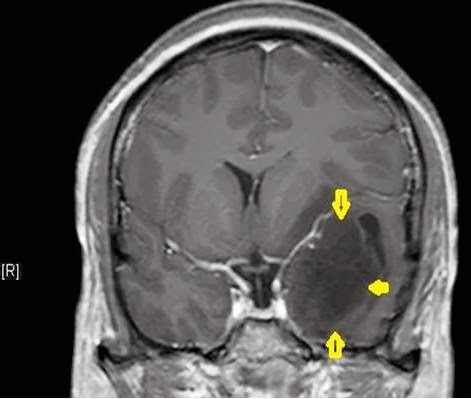
MRI brain also revealed an Arterio-venous malformation in the right temporal lobe(yellow arrows)


Digital Subtraction Angiography showed that the malformation was fed by branches from the middle cerebral artery with veins draining towards the surface and into the depth.


The AV malformation was completely removed by microsurgical technique. During surgery multiple tortuous arterialized veins were fed by right first part of the middle cerebral artery. It was drained by two veins, one over the brain surface and the other towards its lower surface.

Post-operative CT scan of head showed surgical changes and the cavity left after removal of the AVM (yellow dots)


The patient recovered well and was discharged intact on long term anti-epileptic medication.
 
The patient and was advised to get a CT angiogram of the Brain after 3 months of surgery during follow up.


Sunday, February 9, 2014

Limited Dorsal Spinal Rachischisis


A lady aged 28 presented with low backache for 2 years. The pain radiated to both her legs and had increased in intensity for the past 1 year.

She had a skin dimple over her lower back which had been present since birth.

MRI of the spine revealed a fibrous band attached to the skin of the back which then coursed through the spinal cord and was attached to the cord’s coverings at its front. The spinal cord was tethered and low lying, ending 3 vertebral levels below normal.

A diagnosis of Limited Dorsal Spinal Rachischisis was made.

The dermal sinus was excised and the cord de-tethered by microsurgical technique.

Astrocytoma, WHO Grade II

A man aged 29 years presented with episodes of sensation of abnormal taste followed by a fit 6 months later.

He had slight weakness on one side of his face.


The tumor was removed by microsurgical technique and gross total excision was done.


 Biopsy reported it as brain tumor known as an Astrocytoma, WHO Grade II


Monday, January 27, 2014

Trigeminal Neuralgia


This gentleman aged 48 years presented with episodes of excruciating pain over the right side of his
face for 12 years. The pain was brought on by chewing food or shaving and lasted only a few seconds.
An MRI scan revealed a blood vessel indenting the trigeminal nerve i.e. the nerve concerned with sensations over the face.
The patient was treated by “Micro-vascular decompression” in which the nerve was cleared of the offending vessel and Teflon patches placed between them above and below, to prevent further
irritation of the nerve.
The patient was completely relieved of his pain.

Posterior 3rd Ventricle Tumor


A boy aged six presented with episodes of headache and vomiting for about 1 year.

He was drowsy and could not turn his eyes upwards.

MRI of brain revealed a tumor towards the back of the third ventricle (the water system of the brain) with moderate hydrocephalus (dilatation of the water system of the brain).

The tumor was removed by microsurgical technique from the back of the head.

The size of the ventricles of the brain decreased towards normal after surgery.

The patient too improved after surgery and was ambulatory at discharge from hospital.