Pages

Thursday, March 20, 2014

Cerebral Arteriovenous Malformation


A gentleman aged 38 years presented with a 5 years history of a feeling off epigastric discomfort followed by automatic movements lasting for 10 minutes followed by complete recovery. He had no memory of the episode. He was diagnosed as a case of complex partial seizures which were poorly controlled with medication.
 
CT scan of the head showed a lesion in the right temporal lobe of the brain(yellow arrows).


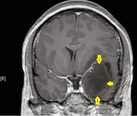
MRI brain also revealed an Arterio-venous malformation in the right temporal lobe(yellow arrows)


Digital Subtraction Angiography showed that the malformation was fed by branches from the middle cerebral artery with veins draining towards the surface and into the depth.


The AV malformation was completely removed by microsurgical technique. During surgery multiple tortuous arterialized veins were fed by right first part of the middle cerebral artery. It was drained by two veins, one over the brain surface and the other towards its lower surface.

Post-operative CT scan of head showed surgical changes and the cavity left after removal of the AVM (yellow dots)


The patient recovered well and was discharged intact on long term anti-epileptic medication.
 
The patient and was advised to get a CT angiogram of the Brain after 3 months of surgery during follow up.


Sunday, February 9, 2014

Limited Dorsal Spinal Rachischisis


A lady aged 28 presented with low backache for 2 years. The pain radiated to both her legs and had increased in intensity for the past 1 year.

She had a skin dimple over her lower back which had been present since birth.

MRI of the spine revealed a fibrous band attached to the skin of the back which then coursed through the spinal cord and was attached to the cord’s coverings at its front. The spinal cord was tethered and low lying, ending 3 vertebral levels below normal.

A diagnosis of Limited Dorsal Spinal Rachischisis was made.

The dermal sinus was excised and the cord de-tethered by microsurgical technique.

Astrocytoma, WHO Grade II

A man aged 29 years presented with episodes of sensation of abnormal taste followed by a fit 6 months later.

He had slight weakness on one side of his face.


The tumor was removed by microsurgical technique and gross total excision was done.


 Biopsy reported it as brain tumor known as an Astrocytoma, WHO Grade II


Monday, January 27, 2014

Trigeminal Neuralgia


This gentleman aged 48 years presented with episodes of excruciating pain over the right side of his
face for 12 years. The pain was brought on by chewing food or shaving and lasted only a few seconds.
An MRI scan revealed a blood vessel indenting the trigeminal nerve i.e. the nerve concerned with sensations over the face.
The patient was treated by “Micro-vascular decompression” in which the nerve was cleared of the offending vessel and Teflon patches placed between them above and below, to prevent further
irritation of the nerve.
The patient was completely relieved of his pain.

Posterior 3rd Ventricle Tumor


A boy aged six presented with episodes of headache and vomiting for about 1 year.

He was drowsy and could not turn his eyes upwards.

MRI of brain revealed a tumor towards the back of the third ventricle (the water system of the brain) with moderate hydrocephalus (dilatation of the water system of the brain).

The tumor was removed by microsurgical technique from the back of the head.

The size of the ventricles of the brain decreased towards normal after surgery.

The patient too improved after surgery and was ambulatory at discharge from hospital.

Monday, October 7, 2013

Basi-Frontal Meningioma

A 45 year old gentleman presented with inability to smell for 20 years and recent onset alteration of memory. MRI Scan of his brain showed a Large Tumour at the base of the brain, behind the forehead – a Basi-Frontal Meningioma.

The tumour was removed completely and the patient recovered nicely. Histopathology (microscopic assessment of the tumour tissue by Neuro-Pathologist) showed a Transitional Meningioma (WHO grade I) – a benign tumour.  

These tumours have a rich blood supply and are prone to bleeding during surgery. The nerves of smell along with centres for memory, behaviour and continence are located in this area of the brain. The nerves of vision as well as blood supply to one’s legs are closely related. Therefore, careful removal of the tumour by microsurgical technique is essential to avoid damaging these structures. 


Saturday, September 28, 2013

Tuberous Sclerosis

A 10 year old boy, third in order of siblings and of normal birth weight, with no complications of pregnancy or child-birth, presented with epileptic fits since childhood along with delayed development and poor performance at school. He also had fleshy growths over his nose and cheeks since 2 years of age.

The epileptic fits involved his right arm during which he would fall unconsciousness followed by a period of confusion before full recovery. The fits occurred during sleep too. Under medication the patient had not suffered fits for the past 2 years.


Even though in fifth standard, he lagged behind in class. However, he was able to look after himself.

An MRI of his brain revealed multiple lesions in the brain with cysts lining the ventricles (the water channels of the brain) and multiple tubers (abnormally formed brain matter) protruding into the ventricles.

An ultrasound of his abdomen revealed polycystic disease of kidneys.

A diagnosis Tuberous Sclerosis was made.


As the patient was free of epileptic fits and none of the tubers in the brain were large enough to cause obstruction of the ventricles no surgical intervention was warranted.

However, as these tubers can transform into brain tumours known as Sub-ependymal Giant Cell Astrocytoma (SEGA) which can obstruct the ventricles (the water system of the brain) causing a rise of   pressure in the brain. They may also cause focal symptoms (weakness of limbs, loss of sensation over the body etc).


The patient should be assessed yearly or earlier in case he deteriorates with a view to surgical intervention to remove the brain tumours if it is required.

However, Tuberous Sclerosis is a multi-system disease which may involve the brain, lungs, heart, kidneys and skin and requires a comprehensive multispecialty team approach to care for these patients.


Case submitted by : A group of Neurosurgeon India

Friday, September 20, 2013

Tumour of the Brainstem ( the part that connects the brain to the spinal cord)


A 3 ½ years child suffered difficulty in walking which progressed to weakness of her right arm and leg ,slurred speech and a squint in her left eye. She was found to have a tumour of the brainstem ( the part that connects the brain to the spinal cord) and was treated with radiotherapy.


The patient improved but deteriorated again about 7 months after radiotherapy. Now she was unable to walk with marked weakness of her right limbs, nearly incoherent speech and a squint of the left eye.


MRI of brain showed a large tumour in the pons (part of the brainstem) on the left side which was bulging towards the 4th ventricle (the water channels of the brain). MR tractography was done to assess the relation of the relay tracts with the tumour which revealed their absence/ destruction in the involved area, so explaining the patient’s symptoms.

Pre-op MR Tractography


The patient’s tumour was removed by microsurgical technique and postoperative scan showed satisfactory removal of the tumour. Postoperatively she showed smooth recovery and was discharged from hospital after she started eating adequate food.
Biopsy showed the tumour to be a malignant tumour (Pontine Glioma - WHO grade III).


                     Post-operative scan


The major advantage of surgery  in patients of tumours of the brainstem is that it reduces its bulk of the tumour and allows one to know the exact nature of the tumour as well. This helps to plan future radiotherapy/ chemo-therapy on the small amount of remaining tumour more effectively thereby translating into improved results. However surgery on the brainstem is very risky as the breathing & heart control centres are there along with the control of all four limbs and the head and neck. So surgery should be done only at centres with full facilities and experienced surgeons.                                                                        

Saturday, October 13, 2012

Orbital Rhabdomyosarcoma

9 years boy
C/o
Proptosis left eye - 3 months
Drooping of left upper eye lid - 2 months
On Examination
         Left ptosis with proptosis
         Fundii normal
         EOM - restricted in superior & lateral direction [left eye]
         Va – Normal
         Vf – Normal in right eye
Pre Op CT Scan

Pre Op MRI

Pre Op MRI

OPERATIVE PROCEDURE:
Left frontal trephine craniotomy, extradural superior orbital approach and microsurgical excision of tumor.
FINDINGS:
Extraconal well defined, non-encapsulated, soft, mild vascular, whitish grey tumor attached to levator muscle.
Complete excision done
Post Op
Left ptosis
Decreased proptosis
Left superior rectus paresis
Subconjuctival hemorrhage
Histopathology


  • Rhabdomyosarcoma - Most common pediatric soft tissue sarcomas  (approximately 50%)
  • Peak age 2- 5 years
  • Male preponderance (1.4:1)
Epidemiology
  • 1/3rd  of RMS patients have other congenital abnormalities
    • GI, GU, CV, CNS
  • Majority cases sporadic; but some associated with genetic conditions
    • Li Fraumeni (p53 mutation)
    • NF 1
    • Beckwith - Wiedemann
Pattern of spread
  • Locally invasive tumor often with pseudocapsule
  • Potential for local spread along fascial or muscle planes, lymphatic extension and hematogenous dissemination
  • Regional lymphatic & hematogenous metastases -15%
Prognostic Factors
  • Histology
  • Stage
    • Primary site (most important)
    • Tumor Size
    • LN involvement
    • Metastatic disease
  • Group
    • Extent of resection
  • Age
    • < 1
    • >10
    • Skull base erosion, CN palsy & Intracranial extension
Histology
  • Gross disease
    • Soft, fleshy tumors with variation in extent of invasion and necrosis
  • IHC stains to ascertain muscle of origin
    • Antidesmin, antivimentin,  anti-muscle specific actin
    • Anti-Myo D
  • Embryonal
    • Most common
    • 60-70% of all childhood RMS
  • Boytroid
  • Spindle cell
  • Alveolar
  • Undifferentiated/pleomorphic
Histology and Survival

Clinical presentation
  • Asymptomatic mass
  • Rapid growing painless mass
  • Mass effect on associated organs and tissues
  • Commonest site orbit > parameningeal sites > other sites
Staging System
Intergroup Rhabdomyosarcoma Study Clinical Grouping Classification


Prognostic Factors
         Orbit
§  favorable prognostic site
§  lymphatic extension/hematogenous metastasis is rare
§  embryonal histology

Management
    • Surgical procedure either excision/biopsy
    • Chemo-radiation
    • Cure rates of  > 90%